Mustafa
Karaça*, Erol Kiliç*, Betül Yazici*,
Sedat Demir* and Jack de la Torre†
*Department
of Neurosurgery, Ihlas Medical Center, Bursa, Turkey
†Department of Pathology, School of
Medicine, University of California - San Diego, La
Jolla, CA, USA
The safety and tolerability of a free radical scavenger with Na+ channel blocking activity (dimethyl sulfoxide (DMSO)) combined with a glycolytic intermediate and high energy substrate (fructose 1,6- disphosphate (FDP)) were assessed in a mostly elderly patient group presenting with acute and subacute ischemic stroke. Eleven patients (average age 65) were given i.v. infusions of DMSO - FDP twice daily for an average of 12 days, while five control patients (average age 63) were given standard therapy. Safety and tolerability were evaluated by clinical adverse effects to drug therapy. Efficacy of DMSO - FDP was assessed by MRI lesion size, by magnetic resonance angiography of ischemic territory, and by a 5-point neurologic recovery scale that rated sensory-motor function and level of consciousness. Results suggest that DMSO - FDP administration is safe, well-tolerated and may be of benefit when given within 12 h after the onset of stroke symptoms. No significant changes in blood pressure, EKG, heart rate or hematology and chemistry profiles, were recorded in any patient receiving DMSO - FDP. Neurologic evaluation at 1, 3 and 6 months after treatments revealed that 7 of 11 (63%) patients given DMSO - FDP achieved 'improved' or 'markedly improved' status while 1 of 5 (20%) standard treated patients showed 'improved' status and only at the 3- month follow-up. This preliminary trial indicates that DMSO - FDP is well tolerated by this group of elderly patients and could be of benefit in reducing neurologic disability after stroke. [Neurol Res 2002; 24: 73 - 80]
Keywords:
Dimethyl sulfoxide; fructose 1,6-diphosphate; ischemic
stroke; neuroprotection; cerebral blood flow; clinical
trial
About 75% of stroke cases involve people over the age of 551. Elderly stroke patients are also more susceptible to neurologic disability and adverse events related to drug treatment following a cerebrovascular insult. Partly for these reasons, aging patients are not usually included in large randomized clinical stroke trials involving investigational drug therapy2.
In the present study, 11 mostly elderly patients ranging in age from 45 to 92 (mean age 65) were randomly selected to receive an investigational drug preparation consisting of dimethyl sulfoxide (DMSO), and fructose 1,6-diphosphate (FDP), following acute ischemic stroke. A second group of five patients received standard treatment.
The main objective of the current trial was to evaluate the safety of repeated doses of sterile, i.v. DMSO - FDP, (product name PHA-56 (Pharma 21, Portland, OR, USA) vs. standard treatment. A second objective was to record the effects of this therapy on post-treatment neurologic and functional outcome at specific time points. Informed consent was obtained from all patients or their legal guardians prior to participation in this study.
The rationale for using combined DMSO - FDP on ischemic stroke patients is based on the clinical and experimental data indicating the beneficial properties of DMSO or FDP monotherapy following brain and cardiac ischemia. Because neither DMSO or FDP alone is able to target the multifactorial metabolic and physiologic abnormalities seen experimentally in animals or clinically in humans, it was considered that their use together might provide improved neuroprotection following ischemic stroke. Neither drug has been used for human stroke before. DMSO has been shown to be a powerful hydroxyl radical scavenger3,4, sodium channel blocker5, microvascular platelet de-aggregator6, and cell membrane stabilizer7,8. Additionally, DMSO has the ability to increase cerebral blood flow (CBF)9-12 when given for a variety of vascular insults and to reduce intracranial pressure in humans following severe head injury13,14. DMSO has been reported to improve neurologic and functional outcome after stroke in animals, including nonhuman primates15-19. DMSO however, is not a substrate for aerobic glycolysis, nor does it reduce ischemic-induced cellular acidosis, affect serum calcium levels or directly increase the synthesis of ATP, the primary energy fuel for all cells, including neurons and glia.
By contrast, administration of FDP has been shown to restore cell energy loss following ischemia or shock by increasing ATP synthesis20. FDP is able to do this because it is an energy substrate booster whose net ATP yield in anaerobic glycolysis is double that of glucose on a per mole basis20. FDP has been shown to reduce the harmful effects of cellular acidosis promoted by lactic acid production following cerebral ischemia21 and to diminish the effects of brain ischemic hypoxia22 possibly by lowering serum calcium levels during the first 24 h after the cerebrovascular insult23. FDP has been shown to enhance energy production by the aerobic glycolysis pathway following ischemia and shock and human clinical studies have shown the beneficial effects of FDP in myocardial infarction20.
Experimental treatment in animals given DMSO - FDP showed this drug combination protected from neuronal damage, sensory-motor deficits and memory loss following cerebrovascular ischemia and traumatic brain injury24,25. This outcome was better than when either agent was given alone24,25. The combination of DMSO - FDP appeared safe and well-tolerated in the animal models tested after 14 weeks of daily administration24. For these reasons, a pilot study using combined DMSO - FDP for the treatment of ischemic stroke in patients was initiated.
A total of 16 patients aged 45 - 92 suffering from acute and subacute cerebral infarction were studied. Patients were entered for this pilot trial from the Ihlas Medical Center (Bursa, Turkey). Median patient age in the DMSO - FDP group was 65 years (six males, five females); five patients (three females, two males) in the standard treatment group had a median age of 63 years. A diagnosis of ischemic stroke was established by clinical signs and symptoms and by MRI of the brain. Patients in the current trial who received DMSO - FDP or standard treatment were moderately to severely impaired. There were eight severely impaired patients in the DMSO - FDP group and two severely impaired in the control group.
Eligible patients who delayed seeking treatment for 6 h or more after the stroke incident, were selected to receive either standard therapy consisting of symptomatic stabilization of vital signs (i.v. fluids, oxygen) and when indicated, osmotic agents, or an intravenous solution composed of dimethyl sulfoxide (DMSO: 560 mg kg-1, 28% solution) and fructose 1,6-di phosphate (FDP: 200 mg kg-1) mixed in 5% dextrose with water, given twice daily for an average of 12 days.
Safety and tolerability to DMSO - FDP were evaluated by recording any clinical adverse effects to drug therapy, by magnetic resonance imaging (MRI) and by laboratory data. Drug efficacy was assessed by a reduction in infarct size using MRI, as well as any mass effect, midline shift or extent of edema, and by magnetic resonance (MR) angiographic evidence of improved perfusion in affected vessels. Neurologic and medical assessments were performed immediately before, during, and after treatments and on 7, 30 and 90 days following therapy. All but one of the 11 experimental drug-treated patients was followed-up for six months after discharge from the hospital. Neurologic assessment included patients' sensory-motor function and level of consciousness. The modified Rankin scale (MRS) was used to predict outcome and a 5-point rating scale adapted from Tazaki et al.26 assessed neurologic recovery (Table 1). All patients were assessed as to their daily living condition as follows: 1, markedly improved; 2, improved; 3, slightly improved; 4, no change; 5, deteriorated or died.
Laboratory and medical examinations were performed before and during treatments at regular intervals. Medical signs were closely monitored and included cardiovascular examination, electrocardiogram, blood pressure, respiratory rate, pulse rate and body temperature. Laboratory tests included complete hematology and chemistry profiles including blood/platelet counts, prothrombin/partial thromboplastin time, blood glucose levels, serum electrolytes, liver/kidney function tests and urianalysis (blood urea nitrogen, plasma glucose, creatinine, electrolytes).
There
were no deaths that could be attributed to any therapy
in this series of patients. Table 1
summarizes the results of the neurologic examination using
the 5-point scale measuring recovery after DMSO - FDP
and standard treatment at 1 and 3 months follow-up in
16 patients presenting with ischemic stroke. No major
changes in the neurological evaluation of patients were
seen between the 3 and 6 month follow-up.
Mean time-to-treatment in the DMSO - FDP group was 8 h in 3 of 11 patients and > 12 h in the remaining eight patients. Of the three patients treated within 6 - 12 h of their stroke, all showed markedly improved or improved status at 1 and 3 months following DMSO - FDP treatment. Of the eight patients who were treated after 12 h, three showed no change from their original pre-treatment status, one showed slight improvement and four attained markedly improved or improved status 3 months after DMSO - FDP administration. Mean time-to-treatment in the five control subjects was 7 h, of these, three patients were slightly improved, one patient improved and one patient remained unchanged after 3 months (Table 1).
Table 1
Eleven
patients treated with PHA-56 (dimethyl sulfoxide + 1,6-fructose
diphosphate) and five control patients given standard
treatment (standard tx) following ischemic stroke
| Patient | Treatment | After 1 month | After 3 months | Tx time (h) |
| ES (90/M) | PHA-56 | Markedly improved | Markedly improved | 6 - 12 |
| FG (62/M) | PHA-56 | Slightly improved | Slightly improved | >48 |
| I U (80/F) | PHA-56 | Unchanged | Unchanged | >48 |
| NT (41/F) | PHA-56 | Improved | Markedly improved | >48 |
| NC (61/M) | PHA-56 | Slightly improved | Improved | >48 |
| HD (59/F) | PHA-56 | Unchanged | Unchanged | >48 |
| GK (80/M) | PHA-56 | Markedly improved | Markedly improved | >48 |
| FO (75/M) | PHA-56 | Markedly improved | Markedly improved | 13-48 |
| EF (60/M) | PHA-56 | Unchanged | Unchanged | >48 |
| AE (62/F) | PHA-56 | Improved | Markedly improved | 6 - 12 |
| IC (63/F) | PHA-56 | Improved | Markedly improved | 6 - 12 |
| HH (74/F) | Standard tx | Unchanged | Slightly improved | 6 - 12 |
| RK (59/F) | Standard tx | Unchanged | Slightly improved | 6 - 12 |
| NA (64/M) | Standard tx | Slightly improved | Improved | 6 - 12 |
| MA (61/M) | Standard tx | Unchanged | Slightly improved | 13 - 48 |
| HK (48/F) | Standard tx | Unchanged | Unchanged | 6 - 12 |
Age and sex for each patient is shown in parentheses. Treatment time (Tx time) after stroke is shown for three time points:1, treatment between 6 - 12 h; 2, treatment between 13 - 48 h; 3, treatment over 48 h. Neurologic evaluation used the Tazaki Neurologic Recovery 5 Point Scale26: 1, markedly improved; 2, improved; 3, slightly improved; 4, unchanged; 5, worsened. Table indicates that 6 (54%) of patients treated with PHA-56 had markedly-improved or improved status 1 month after treatment while no patient with standard treatment attained this neurologic level. At 3 months, 7 (63%) of PHA-56 treated patients and 1 (20%) standard treated patient showed any improvement in their status. Neurologic status did not change significantly in patients followed up for 6 months after treatments.

Figure 1 A: Coronal T2-weighted spin echo image showing ischemic stroke in medial left frontal area invading the thalamus, corpus callosum, cingulate region and surrounding gray matter before treatment (arrows). B: Eight days after twice daily DMSO - FDP i.v. treatment (arrow), there is less gray matter, thalamic and cingulate region involvement. Patient IC, male, age 63
Incidence
and type of side effects were similar for both DMSO
- FDP and standard treated groups. There were no significant
changes in blood pressure, EKG or heart rate in patients
receiving DMSO - FDP. Serum glucose levels monitored
at regular intervals in all patients after treatment
remained within normal limits for the period of observation.
No adverse drug event was observed in any patient given
DMSO - FDP. The combination treatment was well-tolerated
and safe when given i.v. twice a day for an average
of 12 days. Figure 1A shows an MRI
of patient IC immediately prior to treatment with DMSO
- FDP. The lesion extended to the left corpus callosum,
thalamus and cingulate cortex. A second MRI taken 8
days following twice daily i.v. DMSO - FDP (Figure
1B) revealed less anterior gray matter and thalamic
involvement. Figure 2A is an MRI
of patient AE with a large, left posterior cerebral
artery (PCA) territory infarct, lateral mass effect
on the lateral ventricle, left thalamic involvement
and widespread edema of the brain stem. After 11 days
twice daily DMSO - FDP infusions, MRI shows dramatic
reduction of edema, reduced thalamic involvement and
lower signal intensity in gray matter. Magnetic resonance
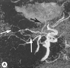
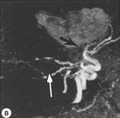
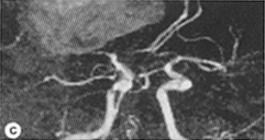
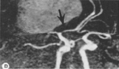
angiogram (MRA) of patient GK (Figure
3B) after a right middle cerebral artery infarct
resulted in a mild mass effect and large hematoma affecting
the basal ganglia territory. MRA of same patient 8 days
after twice daily DMSO - FDP administration showed improved
perfusion in the MCA territory and reduced basal ganglia
involvement (Figure 3A). MRS evaluation
showed 7 of 11 patients treated with DMSO - FDP and
1 of 5 treated with standard treatment had moderate
to no disability (MRS = 0 - 3) after 3 months.
Figure 2

Figure
2 A: T2-weighted SE coronal view MRI of posterior
cerebral artery (PCA) territory infarct before treatment.
White matter changes are seen with lateral mass effect
on lateral ventricle, left thalamus involvement and
widespread edema involving the brainstem (arrows). B:
Eleven days of daily DMSO - FDP i.v. administration,
dramatic reduction of edema and lower signal intensity
with some apparent improvement in gray matter and thalamic
involvement (arrow). Patient AE, male, age 62. See text
for additional information on this patient
Ischemic
stroke is one of the leading causes of death and chronic
disability in the world with elderly individuals showing
the highest prevalence1.
The primary objectives of useful pharmacotherapy in the treatment of stroke should aim to prevent neuronal tissue within the ischemic territory from undergoing further damage, and return, whenever possible, damaged brain cells to normal function. This 'stop and reverse' neuroprotective strategy is dependent on many factors that can influence the outcome of cerebrovascular insults and for that reason, its historical application in stroke has been less than successful in the past. However, one of the 'gold standards' for gauging effective stroke therapy is reduction of lesion size, which includes tissue edema in the ischemic territory27.
Thrombolysis with plasminogen activators has been shown to be effective only within a 3 h window following ischemic stroke symptoms after which these clot busters carry a high risk of intracerebral hemorrhage28. After a considerable number of clinical trial failures, the lack of an effective treatment for ischemic stroke has evoked the sentiment among experts that the probability of discovering a new agent that can successfully treat this condition, appears increasingly distant29. This rather pessimistic view is due in part to the historical pursuit of the 'magic bullet' for brain injury, which subscribes to the concept that a single drug can block or reverse the major pathologic events that unfold during the first 72 h following a stroke. The idea of a stroke 'cocktail' has been suggested in the past30,31 but the actual mixture of two or more effective compounds into one solution that can work synergistically and still attain a level of relative safety, is far from simple since stringent therapeutic and government regulatory processes must be met.
The present combination of dimethyl sulfoxide-fructose 1,6-disphosphate (DMSO - FDP) is an attempt to address critical pathologic events associated with ischemic stroke. These events are widely accepted to be: excessive hydroxyl radical formation leading to oxidative stress32, energy substrate depletion33, inflammatory response34,35, CBF reduction36,37, brain edema37, excitotoxicity38,39, platelet aggregation in the micro-circulation40 and sodium channel activation41.
The combination of DMSO - FDP may target the pathologic formation of cerebral edema, free radicals, inflammation and Na+ channel activation, while improving CBF and energy substrate depletion following an ischemic stroke (Figure 4). For example, DMSO has been shown to be a powerful free radical scavenger3,4,9 with anti-inflammatory and membrane stabilizing activity42,43. DMSO can reduce brain edema and the effects of ischemic stroke in animal models, including non-human primates and in humans9-12,44-46. DMSO additionally increases CBF in a variety of clinical or experimental cerebral insults, possibly as a result of reducing tissue edema, de-aggregating platelet formation and lowering cerebrovascular resistance6,5-19. Drugs that block voltage-dependent Na+ channels are reported to provide strong, neuroprotective activity in animal models of brain ischemia/hypoxia and a number of clinical trials are now in progress to test these class of drugs in ischemic stroke41,37-49. DMSO has been shown to be an effective Na+ channel blocker and this property could partly explain its beneficial effect when administered to the present group of patients5.
  |
  Figure 3: Sagittal view of magnetic resonance angiograph of internal carotid artery territory showing right middle cerebral artery (MCA) infarction with mild mass effect, large hematoma and midline shift involving basal ganglia before treatment (arrows, B, C). Eight days of daily DMSO - FDP infusions reveals no change in hematoma size (as expected), but increased perfusion of the MCA ischemic region with less basal ganglia involvement (arrows, A) and slightly magnified view of MCA flow improvement (arrow, D). Patient GK, female, age 80 |
FDP also has some unusual biological properties that appear relevant in the treatment of ischemic stroke. FDP is a naturally occurring high-energy substrate whose primary function is to participate in the anaerobic glycolytic process to provide cellular energy to the cell. Its administration during ischemic-hypoxia in neonatal rat brain has been shown to improve brain metabolism50 possibly by increasing production of ATP20.
Maintaining cellular ATP levels may prevent or delay many of the negative effects of the biochemical cascade known to occur following cerebral ischemia. Exogenous administration of FDP can restore ATP and oxidative phosphorylation while boosting energy production in the citric acid cycle (aerobic glycolysis) when prolonged hypoperfusion states are present5. FDP also displays free radical scavenging properties20 although not as potent as those observed with DMSO3. Animal experiments from our laboratory and those of others4,9,10,15-23 have shown the effective action of DMSO and of FDP when used alone in CNS insults but it was their combined administration in experimental brain injury using one solution that resulted in markedly improved results over the use of either agent alone24,25. The administration of DMSO - FDP in experimental brain trauma25 and in aging rats subjected to 14 weeks of cerebral hypoperfusion24, has revealed that this drug combination has the ability to 1, protect neurons from mechanical and physiologic injury; 2, reduce sensory-motor deficits following brain damage; 3, restore visuospatial memory loss during chronic brain ischemia in aging rats; 4, restore loss of protein synthesis; and 5, reduce astroglial reactivity24,25. In addition, a recent study has shown that DMSO can prevent glutamate-induced excitotoxic death in cell cultured hippocampal neurons and prevent excessive calcium influx into these cells52, an extremely critical mechanism that is central to neuroprotective action.
Figure 4

Figure
4: Sketch of pathologic events potentially targeted
by dimethyl sulfoxide+ fructose 1,6-diphosphate (PHA-56)
following ischemic stroke. Clinical and experimental
findings (see text for details) have reported dimethyl
sulfoxide lowering brain edema, scavenging free radicals,
blocking sodium channel activation, reducing inflammation
and increasing cerebral perfusion, while fructose 1,6-diphosphate
is reported to improve aerobic glycolysis and increase
ATP synthesis in the presence of cerebral ischemia
Although the present pilot study contained a relatively small number of experimental and control subjects, some general observations seem appropriate with respect to the safety and tolerability of continued DMSO - FDP infusions over a 2-week period.
DMSO - FDP appears safe and well-tolerated at the doses used and no adverse drug reaction was recorded in any patient during the 3-month observation period or at the 6-month follow-up, when patients were given a final assessment. In addition, DMSO - FDP may have benefited some of the patients in this study when compared to standard treated subjects (Table 1). For example, most of the patients neurologically rated as markedly improved/improved, showed a reduced lesion size several days following treatment (Figures 1 and 2), but lesion size appeared generally unaltered in the control patients. Although reduced lesion size after ischemic stroke does not always correlate with the pathologic examination, in most cases, when it does not, it may indicate that tissue edema has been reduced around the infarcted vessel. Vessel patency and revascularization around the ischemic lesion as assessed by magnetic resonance angiography, appeared improved in some patients within 1 - 2 weeks after receiving DMSO - FDP (Figure 3). Again, this phenomenon could have occurred by chance although none of the standard treated control patients showed such marked reperfusion activity.
In DMSO - FDP treated patients, 54% showed markedly improved or improved status 1 month after treatment compared to 0 of 5 control-treated patients. At the 3-month follow-up, 7 of 11 patients (63%) treated with DMSO - FDP showed markedly improved or improved status while 1 of 5 patients (20%) attained this neurologic level for the same time period.
Neurologic reduction of deficits in the DMSO - FDP treated patients included significant improvement of sensation, motor strength, higher cortical function (speech, alertness, comprehension) and ambulation in follow-ups conducted at 30 and 90 days after discharge from the hospital.
Significant motor improvement was recorded in 54% of the patients 1 month after receiving DMSO - FDP, and this was especially evident in the three subjects treated within 12 h after their stroke symptoms appeared. This compares with 20% of patients with minor motor improvement in the standard treated controls after 1 month. Since some of the DMSO - FDP patients improved their neurologic status even when treatment was applied 13 h after their stroke symptoms first appeared (5 of 11), it is suggested that DMSO - FDP treatment may have neuroprotective qualities. In addition, since the average age of the seven patients achieving a markedly improved or improved status after 3 months was 69 years, drug efficacy did not appear to be negatively correlated with advancing age in this population group. This is an encouraging finding that merits further examination of DMSO - FDP activity on a larger population group particularly since most major stroke trials generally exclude the elderly as study subjects3.
In summary, DMSO - FDP was well-tolerated when given twice daily for an average of 12 days, to mostly elderly subjects following ischemic stroke. Moreover, DMSO - FDP could have provided neuroprotective effects to some of the patients in this small population group during a 3- and 6-month follow-up. While no conclusions can be drawn from these observational findings, the use of DMSO - FDP for ischemic stroke needs to be studied in a large, randomized trial to test the merit of this drug combination under a more control led setting.
The
authors are grateful to Pharma 21 (Portland, OR, USA)
for generous supplies of pharmaceutical grade PHA-56 (dimethyl
sulfoxide-fructose 1,6-diphosphate). The authors wish
to thank Dr Blaine Hart, University of New Mexico, for
helpful comments on the neuroradiologic images.
From
Neurological Research, 2002, Volume 24, January
Correspondence and reprint requests to: JC de la Torre,
MD, PhD, UCSD-Pathology, 1363 Shinly, Suite 100, Escondido,
California 92026, USA. (jdelator@nctimes.net)
Accepted for publication June 2001
Copyright © 2002 Forefront Publishing Group
0161-6412/02/010073-08
© 2001-2022 All rights reserved